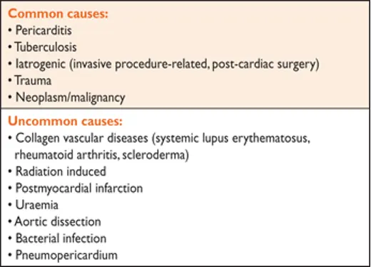
Cardiac tamponade is a life-threatening, slow or rapid compression of the heart due to the pericardial accumulation of fluid, pus, blood, clots or gas as a result of inflammation, trauma, rupture of the heart or aortic dissection.
- H&P
- Clinical signs in a patient with cardiac tamponade include tachycardia, hypotension, pulsus paradoxus, raised jugular venous pressure, muffled heart sounds
- ECG:
- low-voltage
- electrical alternans
- May show signs of pericarditis
- Chest imaging (e.g., CXR) with enlarged cardiac silhouette
- Classic signs include Beck’s triad
- neck vein distension with elevated JVP,
- pulsus paradoxus,
- diminished heart sounds
- Pericardial friction rub can be heard if concomitant pericarditis
- Fun fact: cath will show equilibration of average diastolic pressure and characteristic respiratory reciprocation of cardiac pressures, i.e. an inspiratory increase on the right and a concomitant decrease on the left---the proximate cause of pulsus paradoxus.
- Except in low-pressure tamponade, diastolic pressures throughout the heart are usually in the range of 15-30 mmHg.
Diagnosis
Echo in Cardiac Tamponade
- Echo is the single most useful diagnostic tool to identify pericardial effusion and estimate its size, location and degree of hemodynamic impact
- Early diastolic collapse of RV
- Late diastolic collapse of RA
- abnormal ventricular septal motion
- exaggerated respiratory variability (>25%) in mitral inflow velocity
- > 40% of tricuspid inflow?
- inspiratory decrease and expiratory increase in pulmonary vein diastolic forward flow,
- respiratory variation in ventricular chamber size,
- aortic outflow velocity (echocardiographic pulsus paradoxus) and
- inferior vena cava plethora
Waveforms
- RAP waveform has an attenuated/absent Y-descent
- Occurs d/t diastolic equalization of pressures in the RA and RV + lack of effective flow across the TV in early ventricular diastole
- Can also see equalization of mean RA, RV, and PA diastolic pressures and mean PCWP